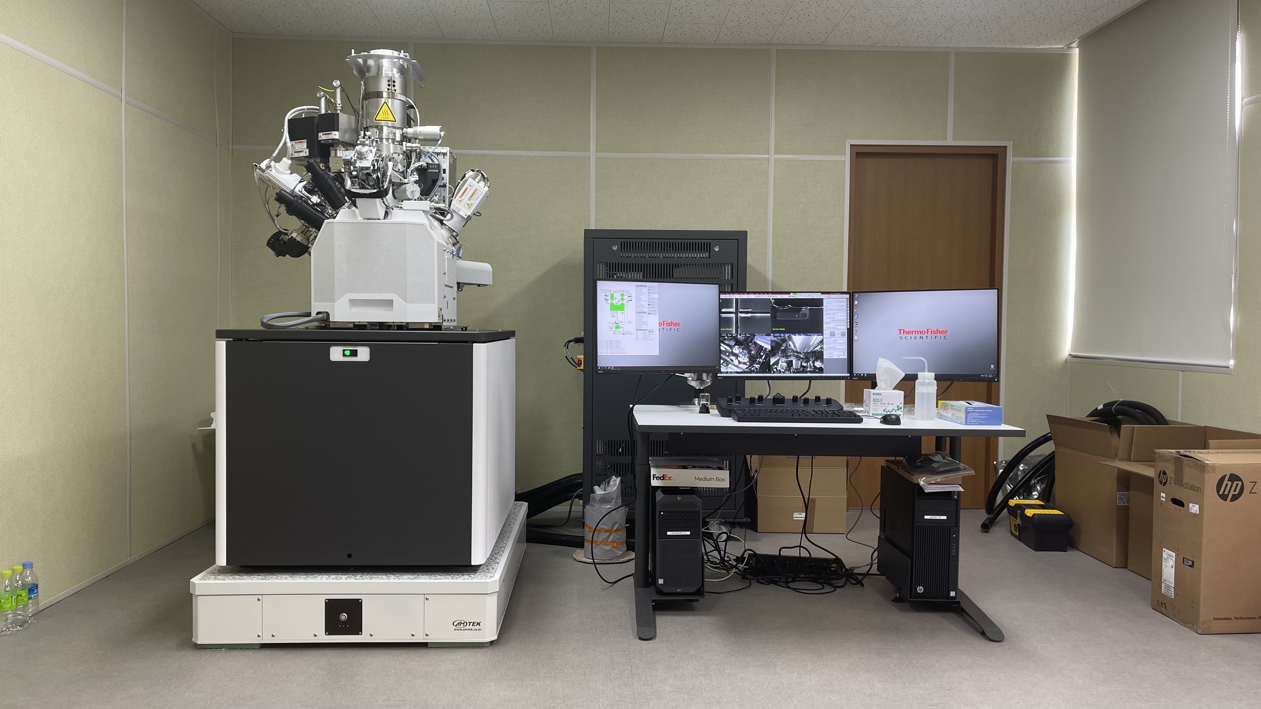
공동활용장비
- Multifunctional Focused Ion Beam System
- 다기능 집속이온빔 시스템
- i-Tube No. 2109-A-0379
- NTIS No NFEC-2021-10-273675
- 설치기관 부경대학교산학협력단
- 주소 부산 남구 용당동 부경대학교용당캠퍼스
- 담당자 권용민 (T. )
- 매뉴얼
-
온라인* 본 장비는 온라인 예약이 불가하오니 장비사용 예약과 관련해서는
예약가능여부
장비 보유기관에 직접 문의 주시기 바랍니다. (장비 예약은 Zeus 시스템에서 회원가입후 예약가능)
장비정보
-
제작사
Thermo Fisher Scientific
-
모델 명
Helios 5 CX
내용연수10년
-
구분
주장비
용도분석
-
표준 분류
광학·전자 영상장비 > 현미경 > 달리 분류되지 않는 현미경
-
취득일
2021-09-29
취득금액931,000,000원
- 인증정보
- 기능
- 장비 상세설명
이용안내
-
사용형태
기관의뢰
설치형태고정형
-
사용료 형태
시간별
장비 사용료300,000원
장비설명
- 본 장비는 박막, powder, Bulk 등 다양한 형태에서 시료의 초미세 형상, 원자 수준의 구조분석 및 화학분석등에서 요구되는 나노구조분석을 제공하는 전자현미경임.
- Nano structure manipulation
- 3D를 활용한 다양한 재료의 성분 및 결정 분석으로 정밀한 hardware 및 자동화된 software, 3D Reconstruction program의 적용으로 다양한 3D 분석이 가능함.
- 자동화 된 가공 및 focus 기능을 가진 Auto Slice & View G3, 3D Visualization program 인 Avizo를 활용하여 3D Tomography, 3D EDS, 3D EBSD 등의 분석이 가능하여, 우수한 데이터를 확보함으로써 첨단소재의 개발 지원이 가능함
- 최고 수준의 이온빔 정밀도를 가지고 있어, 수 나노 수준의 이온빔 milling 및 material deposition, Nano-builder를 활용하여 CAD파일을 직접 불러와서 작업이 가능함
- 복잡한 형태의 나노 구조물 및 pattern 도 쉽게 제작이 가능하여 추가적인 반도체 공정을 거치지 않고도 Lab 수준의 prototypes이 가능하며 보다 다양하고 많은 실험을 편리하게 진행 가능함
장비 구성 및 성능
1. FIB basic unit
○ Electron optics
– Electron Beam Resolution
• 0.6 nm at 15kV ~ 2kV landing energy or better
• 1.0 nm at 1kV or better
– Landing energy : 20 V to 30 kV or better
– Beam current : 0.8pA~ 100nA or better
○ Ion optics
– Ion Beam Resolution
• 2.5 nm at 30kV using selective edge method
– Accelerating voltage : 500 V to 30 kV
– Beam current : 0.1pA to 100nA (15 position aperture strip)
– Liquid Gallium ion emitter with two-stage differential pumping
○ Scanning system
High-resolution digital scanning engine controlled from the user interface
– Resolution
• 512x442, 1024x884, 2048x1768, 4096x3536 pixels (conventional)
• 768x512, 1536x1024, 3072x2048, 6144x4096 pixels (wide screen)
– Minimum dwell time: 25 ns / pixel
– Electronic scan rotation by n x 360 degrees
○ Vacuum system
This FIB system must have complete oil free vacuum system
– 1 x 210l/s turbo molecular drag pump
– 1 x PVP (dry pump)
– 4 x IGP (total for electron column & ion column)
– Chamber vacuum: < 2.6 x 10⁻⁶ mbar
○ Chamber
– E-beam and I-beam coincidence point at analytical WD
– Chamber inner size 379mm(Ø) or bigger
– 21 ports or bigger
2. NanoManipulator system
○ Embedded SW controlled sample lift out manipulator system and rotation
- Drift : <50nm / min
- Smallest step size : 50nm
- True Z movement over 5um move :<500nm
- Vibration : <15nm
- Omnidirectional repeatability : <±150nm
- Motorized Rotation
3. Stage system
- X, Y: 110mm or bigger
- Z: 65 mm motorized or bigger
- Tilt : -15° ~ + 90° or bigger
- Rotation : n×360°
- X, Y repeatability 1µm
4. Detectors
- In-lens SE and BSE detector
- Everhart-Thornley Secondary electron detector
- In-column detectors: Low and high energy loss detection
- IR Camera view in the chamber
- Chamber integrated Navigation camera
- Retractable solid state Backscatter detector
- Plasma cleaner system(software operation in UI)
5. Gas injection system
- Platinum and carbon GIS system
6.SDD EDS 60M (129 eV)
- EDS detector for small dual beam chamber SEM
- Active area of 60 mm2 and 129eV energy resolution at Mnk-alpha
- Norvar window with proprietary evacuated tube design for detection sensitivity to Be
- Analyzer electronics with up to 1,000,000 x-ray input counts per second and 300,000 x-ray output counts per second.
- Motorized slide for software controllable insertion / retraction.
7. Accessories
- Air compressor
- EMI UPS system
- Water Chiller system
8. System control & software
- 64-bit Graphical User Interface with Windows operation system, keyboard, optical mouse
- Microscope Control Computer
• Intel Workstation Processor
• 2.66 GHz or better- 500 GB system hard drive
• 12 GB RAM- Firewire and Ethernet support
- Three 24 inch LCD displays, WUXGA 1920 x 1200
- Support PC with Windows7 & software controller share one keyboard and mouse
- The stage can be controlled through the user interface


