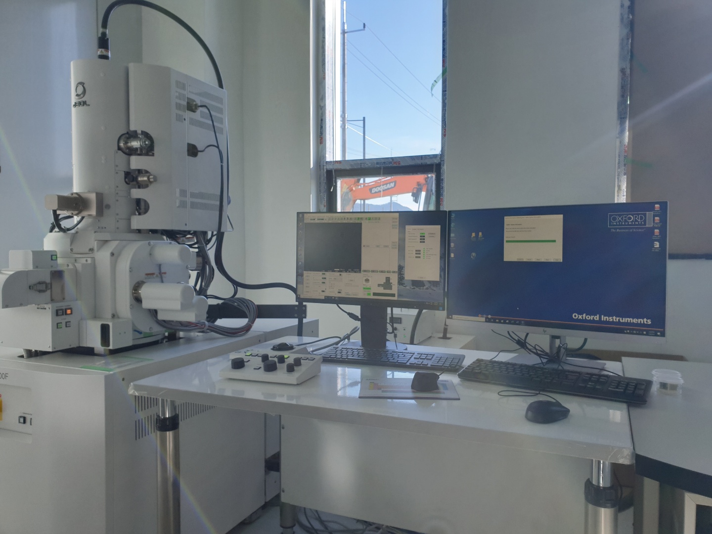
공동활용장비
- Microstructure analysis system for C Composite
- 탄소소재 미세 구조분석 시스템
- i-Tube No. 2101-A-0237
- NTIS No NFEC-2021-04-270017
- 설치기관 (재)경북하이브리드부품연구원
- 주소 경북 영천시 괴연동 36
- 담당자 윤원호 (T. 053-582-3040 )
- 매뉴얼
-
온라인* 본 장비는 온라인 예약이 불가하오니 장비사용 예약과 관련해서는
예약가능여부
장비 보유기관에 직접 문의 주시기 바랍니다. (장비 예약은 Zeus 시스템에서 회원가입후 예약가능)
장비정보
-
제작사
Jeol
-
모델 명
JSM-7900F
내용연수10년
-
구분
주장비
용도분석
-
표준 분류
광학·전자 영상장비 > 현미경 > 주사전자현미경
-
취득일
2021-01-11
취득금액980,489,940원
- 인증정보
- 기능
- 장비 상세설명
이용안내
-
사용형태
기관의뢰
설치형태고정형
-
사용료 형태
시간별
장비 사용료55,000원
장비설명
탄소 소재 미세구조분석 시스템은 일반 광학현미경으로는 관찰이 어려운 탄소 소재의 기공, 함침 적정성을 관찰할 수 있는 복합소재 미세영역을 고분해, 고배율(최저 25배에서 최고 100만배까지)로 확대하여 표면, 단면 구조 및 형태를 확인 가능한 기초 분석 장비
장비 구성 및 성능
장비의 구성(Configurations of Goods)
1. 주장비
1) FE-SEM main body 1 set
2) 5 Axis Motor Drive stage 1 set
3) Specimen Exchange Chamber 1 set
4) Personal Computer with 24inch LCD monitor 1 set
5) UPS for FE-Gun 1 set
6) In column Electron detector system with ACL 1 set
7) Super high resolution bias deceleration system 1 set
8) Auto beam alignment software 1 set
9) Retractable BSE detector 1 set
10) Color image Stage Navigation System 1 set
11) IR Chamber Camera 1 set
12) Embedded type anti-vibration isolation system 1 set
13) 5.2cm bulk holder 1 set
14) Cross section multi specimen holder 1 set
15) Operation Table 1 set
16) Energy Dispersive X-ray Spectrometer 1 set
17) Low damage Cross section system 1 set
18) Automatic magnetron sputter coater 1 set
-. Target : Pt & Au
19) Automatic carbon coater system 1 set
20) Consumable parts
-. Aperture 2 ea
-. Vacuum Grease 1 ea
-. Specimen mount, 12.5Dx5H, 3 ca
-. Specimen mount, 25.4Dx5H, 3 ca
-. Adhesive Tape(carbon), 20M 3 ca
2. 보조장비
1) Cooling Water Circulator for FE-SEM 1 set
2) A.V.R for FE-SEM 1 set
성능 및 규격(Performance and Specification)
1. Performance
1) Image Resolution : 0.7nm guaranteed at 15kV or less
0.8nm guaranteed at 1kV or less
1.0nm guaranteed at 0.5kV or less
2) Magnification : x25 to x1,000,000 on photo mag. or more
3) Image types : Secondary electron image
Backscattered electron image
SE and BSE mixed image
(with filtering system)
4) Accelerating Voltage : 0.02 to 30kV or more
5) Probe current : A few pA to 400nA or more
2. Electron Optical System
1) Lens control method : New electron optical control mechanism.
Synchronized system which control the electron gun and optical system.
2) Electron Gun : In lens Field Emission-Gun
3) Lens System :
-. Observation mode : SEM, LDF GBSH or more
-. Condenser lens (C.L) : Electromagnetic 2-stage lens
-. Aperture angle control lens (ACL) :
Electromagnetic lens
-. Objective lens (O.L) : Hybrid strong-excitation conical objective lens/ Compound type
(electrostatic/electromagnetic filed superposed OL)
-. OL aperture : Click-stop type, 4 step or more
3. Specimen stage
1) Type : Computer controlled 5-axis motor stage
2) Movements : X direction : 70mm or more
Y direction : 50mm or more
Z direction : 39mm or more
Tilt : -5˚ to +70˚ or more
Rotation : 360˚ endless (Motor driven)
3) Specimen exchange : Single touch chucking by Airlock
chamber door open possible.
4) Specimen exchange chamber :
Specimen size : 100mm(dia.)x40mm(h) or more
Isolation valve : Automatic control
5) Analytical condition EDS and EBSD analysis should be possible at the same time.
3.0nm or less resolution at analytical condition. (5kV, 5nA, WD10mm)
6) Beam deceleration system Max. bias voltage : 5kV or more
Min. observation condition : 20V or less
4. Electron detection system
1) Lower detector : E-T type detector or more
2) Upper detector : E-T type detector with electron filter or more
3) BSE Detector : One click auto retractable type detector below OL
Image resolution : 1.5nm at 15kV, WD 4mm or less
Observable WD : 2.0 to 15mm
5. Scanning&Display System
1) SEM control system PC : PC/AT compatible
RAM : 8GB or more
OS : Windows10 professional or more
24 inch LCD monitor or more
2) Scan and display mode : Full-frame display
Direct magnification display
Selected-area scan
CF scan
Dual display
Quad split display or more
3) Scan speed : speeds selectable system from 12speed or more
6. Automatic functions : Automatic focus combination with ACB
Automatic stigmator combination with ACB
Automatic contrast and brightness (ACB)
7. Evacuation System
1) Gun chamber & intermediate chamber :
Ultrahigh vacuum dry evacuation system with ion pumps
2) Specimen chamber : Dry evacuation system using TMP
3) Dry nitrogen gas connector : Included. Used for vacuum release
3) Ultimate pressure
-. Gun Chamber : 10-7Pa or less
-. Specimen Chamber : 10-4Pa or less
4) Vacuum Pumps : Two Ion Pumps or more
One TMP Pump or more
One RP with fore-line trap or more
8. Low damage cross section system for beam sensitive specimen.
1) Beam damage reduce function :
Fine milling & Intermittent milling or more
2) Specimen swing function :
Automatic specimen swing of ±30° during milling.
3) Milling speed : 500um/ hour or more
4) Ion beam source : Ar ion beam
5) Ion beam diameter : 500um or more
6) Ion accelerating voltage : Max 8kV or more
7) Holder system : Rotation flat milling holder
Carbon coating holder
9. Energy Dispersive X-ray Spectrometer
1) EDS System
-. 64 bit Analysis Software with Dual image capture system
-. Quantitative analysis results overlaid in spectrum viewer
-. Quantitative analysis can be achieved automatically with real time
-. Single crystal LN2 Free SDD, 40mm2or larger
-. Energy resolution ≤127eV at 130,000cps or better
-. Resolution guaranteed in accordance with ISO15632:2002
2) Application Software & Optional item
-. Live image acquisition & X-Ray Mapping
-. Live spectrum
-. Live trace
-. Data saving
-. Nano Analysis system
-. Smart map for X-ray mapping & Linescanning
-. Color elementary mapping
-. Realtime Quantitative analysis
-. Beam Drift Correction


